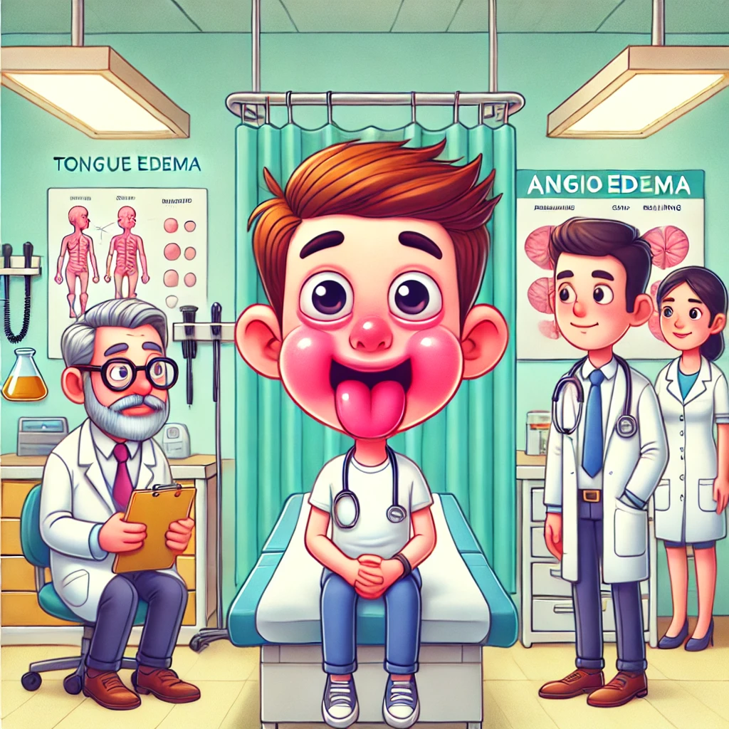
Angioedema is a complex clinical condition characterized by rapid subcutaneous or submucosal swelling. Its diverse etiologies, underlying mechanisms, and varied presentations make it a significant diagnostic challenge. Accurate differentiation between types and their mimics is essential for effective management and preventing serious complications. This blog post provides a comprehensive guide to understanding angioedema, its mimics, and a systematic diagnostic approach.
Understanding Angioedema: Mechanisms and Clinical Features
Angioedema can be categorized based on its underlying mediators—histamine, leukotrienes, and kinins—each with distinct pathophysiological and clinical characteristics.
- Histamine-Mediated Angioedema
• Mechanism: Triggered by IgE-mediated mast cell degranulation.
• Clinical Features:
o Rapid onset (minutes to hours).
o Frequently accompanied by urticaria (wheals or hives) and pruritus.
o Responds well to antihistamines and corticosteroids.
• Common Triggers: Foods (e.g., nuts, shellfish), medications (e.g., antibiotics), insect stings, and latex. - Leukotriene-Mediated Angioedema
• Mechanism: Caused by COX-1 inhibition, leading to leukotriene pathway activation. Commonly associated with NSAID or aspirin use.
• Clinical Features:
o Often presents with urticaria but shows only partial response to antihistamines.
o Symptoms may persist for several days.
• Key Consideration: Frequently seen in patients with aspirin-exacerbated respiratory disease (AERD). - Kinin-Mediated Angioedema
• Mechanism: Driven by bradykinin excess, kinin-mediated angioedema is seen in hereditary angioedema (HAE), acquired angioedema (AAE), and drug-induced cases.
• Clinical Features:
o No true urticaria. Instead, erythema marginatum (a non-itchy, ring-like rash) may be seen in HAE.
o Unresponsive to antihistamines or corticosteroids.
o May involve visceral organs, causing abdominal pain or airway obstruction.
• Subtypes:
o Hereditary Angioedema (HAE):
Caused by C1 inhibitor (C1-INH) deficiency or dysfunction.
In cases of HAE with normal C1-INH, genetic mutations (e.g., Factor XII) are often implicated.
Clinical Note: HAE is not associated with urticaria but may present with erythema marginatum.
o Acquired Angioedema (AAE):
Associated with autoantibodies to C1-INH, leading to immune complex formation and complement activation.
Key Difference: Unlike HAE, AAE may result in urticarial vasculitis, presenting as a true urticaria-like rash due to immune complex deposition.
o Drug-Induced Angioedema:
Often linked to ACE inhibitors, DPP-4 inhibitors, and spironolactone.
Clinical Note: Drug-induced cases lack urticaria and present with isolated lip, tongue, or throat swelling.
Angioedema Mimics: Diagnostic Challenges
Angioedema can mimic other conditions, making accurate diagnosis challenging. Below are key mimics and their distinguishing features:
- Infectious Mimics
• Gnathostomiasis:
o Key Features: Migratory subcutaneous swelling, eosinophilia, and travel history to endemic areas (e.g., Southeast Asia, Latin America).
o Clue: History of consuming raw or undercooked freshwater fish. - Autoimmune and Inflammatory Mimics
• Dermatomyositis:
o Key Features: Facial edema with or without muscle weakness. Elevated muscle enzymes (e.g., CPK) or autoimmune markers (e.g., ANA) confirm the diagnosis.
o Clue: Look for subtle signs like heliotrope rash or Gottron’s papules. - Vascular Mimics
• Superior Vena Cava (SVC) Obstruction:
o Key Features: Morning facial swelling, dyspnea, and neck vein prominence. Imaging (e.g., CT angiography) is diagnostic.
o Clue: Swelling worsens when lying down. - Granulomatous Mimics
• Granulomatous Cheilitis (Melkersson-Rosenthal Syndrome):
o Key Features: Recurrent lip swelling progressing to persistent edema. Biopsy reveals granulomatous inflammation.
o Clue: Look for fissured tongue or facial nerve palsy. - Allergic Mimics with Systemic Features
• DRESS Syndrome (Drug Reaction with Eosinophilia and Systemic Symptoms):
o Scenario: A patient with facial redness and swelling following antibiotic use is initially diagnosed with food-induced allergic angioedema. Days later, they develop a rash, fever, and elevated liver enzymes, confirming DRESS syndrome.
o Key Features: Drug exposure history, systemic involvement (fever, rash, organ dysfunction), eosinophilia, and elevated liver enzymes.
o Clue: A high index of suspicion is required in patients with recent drug exposure and systemic symptoms. - Rare Syndromes
• Mikulicz Syndrome (IgG4-Related Disease):
o Scenario: A middle-aged woman presents with episodic upper eyelid swelling initially thought to be angioedema. Biopsy reveals IgG4-positive plasma cell infiltration, consistent with Mikulicz syndrome.
o Key Features: Recurrent swelling of salivary and lacrimal glands, systemic symptoms, and biopsy findings of IgG4-positive plasma cells.
o Clue: Consider in patients with persistent or recurrent swelling and systemic involvement.
• Gleich Syndrome (Episodic Angioedema with Eosinophilia):
o Scenario: A patient with recurrent episodes of edema, eosinophilia, and sudden weight gain is misdiagnosed with idiopathic angioedema. Eosinophil activation confirms Gleich syndrome.
o Key Features: Episodic edema, peripheral eosinophilia, weight gain, and elevated IgM levels.
o Clue: Look for cyclical patterns of swelling with associated eosinophilia. - Hormonal Causes
• Progesterone-Sensitive Angioedema:
o Key Features: Cyclical swelling linked to the luteal phase, with improvement post-menstruation.
o Clue: Symptom relief with ovulation suppression confirms the diagnosis. - Drug-Induced Angioedema
• Key Features: Sudden lip or tongue swelling without urticaria. Common culprits include ACE inhibitors, DPP-4 inhibitors, and spironolactone.
• Clue: Onset may be delayed (e.g., months to years after starting ACE inhibitors).
Practical Diagnostic Approach
A systematic approach is essential for accurate diagnosis and management:
Comprehensive History:
o Triggers: Medications (e.g., ACE inhibitors, NSAIDs), dietary habits (e.g., raw fish), and travel history.
o Cyclic Patterns: In female patients, assess for hormonal causes like progesterone sensitivity.
o Systemic Symptoms: Fever, rash, weakness, or morning-dependent swelling.
Targeted Examination:
o Look for subtle signs of SVC obstruction, erythema marginatum, or muscle weakness.
o Differentiate between urticaria and non-urticarial rashes.
Laboratory and Imaging:
o C1 Inhibitor Levels and Function: Essential for diagnosing HAE and AAE.
o Eosinophil Count: Suggests parasitic, allergic, or rare syndromes like DRESS or Gleich syndrome.
o Biopsy: Indicated for granulomatous or autoimmune mimics, as well as IgG4-related diseases.
o Imaging: CT or MRI for suspected SVC obstruction or visceral involvement.
Conclusion
Angioedema is a multifaceted condition requiring a nuanced understanding of its underlying mechanisms and mimics. By recognizing the distinct pathways—histamine, leukotriene, and kinin-mediated—and systematically evaluating potential mimics, healthcare professionals can ensure accurate diagnosis and optimal patient care. A thorough history, targeted examination, and appropriate diagnostic testing are the cornerstones of effective management.
Key Takeaways
• Angioedema can be mediated by histamine, leukotrienes, or kinins, each requiring specific management strategies.
• Mimics such as gnathostomiasis, dermatomyositis, DRESS syndrome, and rare syndromes like Mikulicz and Gleich syndromes must be considered in the differential diagnosis.
• A systematic diagnostic approach, including detailed history, examination, and targeted testing, is crucial for accurate diagnosis and treatment.


